4. EARLY VISION LOSS DUE TO RUBEOLA OR A SYNDROME, IN HEARING AND HEARING IMPAIRED CHILDREN
The typical feature of this group of children that makes them different from the Usher group, is the presence of dual sensory impairment from birth. The often severe communication problem is present during the first year when its effect on all areas of development is strong.
Assessment of vision of these children is most important as early as possible during the first year. It should be repeated at regular intervals to guide the nursing staff, therapists and parents in using vision optimally at the same time as all other communication and learning techniques are used without prejudice.
We often picture these children as severely brain damaged children but some of them have normal development of brain functions despite severe sensory deprivation. It is important NOT to make a diagnosis of developmental delay if communication does not function. Communication is a two way street, often it is the adult tester who is the more disabled one in communication with the child.
Typical features of these children are microphthalmia (=small eye), colobomas (=a part missing) of the retina, iris and/or the optic nerve, cataracts of different kind, corneal cloudiness and deviations in the structure of the entire anterior chamber of the eye. In order to understand these numerous deviations from normal development, a short summary of the development of the eye may be helpful.
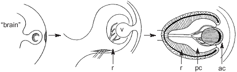
Growth of the eye. r = retina, v = lens vesicle, pc = posterior chamber, ac = anterior chamber.
The development of the eye begins when a finger like structure grows from the front surface of the neural tube in the 4 mm long embryo. An indentation appears on the end surface of this extension, it becomes asymmetric to form a furrow on the side that later will become the lower part of the eye. Through this furrow vessels grow into the space inside the bowl like structure that will become the retina (r). The vessels do not grow into the retina yet but they grow into the tissue inside the retinal bowl. They disappear later when the gelatinous vitreous is formed in the posterior chamber (pc). Some remnants of these vessels may be visible on the optic disc in normal eyes.
The furrow through which the vessels grow into the eye, closes normally. The vessels are then inside the optic nerve and the retina is continuous. If the furrow does not close, it may stay open at different locations, either in the anterior part or in the posterior part of the eye or in both. If closure does not occur in the front part of the eye, there is a defect in the lower part of the retina and choroid, called coloboma. There is often also a defect in the lower part of the iris, producing a "keyhole pupil" . If the closure does not occur in the back part of the eye, the coloboma is in the retina and may extend into the optic nerve. In such a case there is no optic disc but a funnel shaped pit in its place. Since the coloboma is in the lower part of the eye, the corresponding visual field defect is in the upper part of the visual field.
At the same time as the retina develops at the back of the eye, its front edge induces the development of the lens. This is first a fluid filled vesicle (v) or bubble, formed by one cell layer. Then cells in its back surface start to grow to from dense, regularly placed lens fibres. In front of the lens the tissue undergoes cleavage and the anterior chamber (ac) and the cornea (c) are formed. Then iris tissue grows between the lens and the cornea. At this time the ciliary body and the anterior chamber angle also develop.
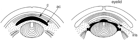
Disturbance in the development of the anterior parts of the eye may occur at different stages and thus interfere with different structures. The lens may not separate from the back surface of the cornea, as in normal development. There may be cataractous changes in the lens, the iris may be missing totally (=aniridia) or parts of it may not have developed. The pupil may be displaced or have an unusual shape. The anterior chamber angle may not have developed the sieve like structures through which the intraocular fluid flows out of the eye and glaucoma may ensue. The cornea may be smaller in diameter than normal (=microcornea) or the whole eye may be smaller than normal (=microphthalmia) . A microphthalmic eye may have fairly good function but is often severely defective because of irregular refractive errors, opacities (=cloudiness) of the cornea and/or the lens and colobomatous changes in the retina and the optic nerve.
Since these malformations are an important cause of vision impairment , it is good to have a description of the structural changes with a draft drawing depicting them. A drawing often makes it possible to understand the effect of the structural changes on the function of the eye. Equally important is to know if there is hypoplasia of the optic nerve, a smaller than normal optic nerve.
Large refractive errors and the need of glasses are common in the group of children with colobomas. Microphthalmic children may have high refractive errors. Prematurely born children with colobomas may also have retinopathy of prematurity and changes in the visual pathways due to periventricular leucomalacia (= changes in the white substance next to the brain ventricles, fluid filled spaces in the brain). Since the motor pathways are located close to the ventricles, these children may have obvious motor problems or their motor defects are so mild that they remain undiagnosed until the assessment of the visual pathways leads to a more careful neurologic examination when minor disturbances in the motor functions may be identified. For the planning of sports and other physical activities it is good to learn even about these minor motor problems.
Auditory impairment and disability and communication problems vary as much as does the vision impairment and disability. It is essential to become familiar with the child's communication techniques and level when starting to plan the assessment of visual function. In the beginning of the assessment the possibility of loss of the upper visual field in children with colobomas is kept in mind and measurement of the extent of the visual field is one of the first tests. The next test is to find out the sphere for visual communication by using the Hiding Heidi test. By testing first at the usual communication distance it is possible to quickly measure at which contrast the child can perceive the Heidi face at different distances.
In visual communication with hearing impaired children with colobomas, the same rules apply as they do for communication with deaf Usher children except that signs that start high up may need to be avoided or the child is guided by the signer's gaze to look up before the gesture is made.
Measurement of visual acuity, reading acuity, contrast sensitivity and colour vision are usually easy once the communication is fluent. Problems of visual adaptation are rare but worth observation at both high and low luminance levels. Measurement of visual field is done either with perimetry, campimetry or using a confrontation technique, depending on the communication level of the child.
If the child has poor contrast sensitivity, facial expressions may appear so blurred that the structure and content of facial expressions is not well known by the child. Heidi Expressions Game can be used to make the child aware of the structure of different expressions. The use of the Heidi expressions-cards can be combined with tactile exploring of facial expressions and drawing the different expressions. When the expressions have been learned at high contrast, then the low contrast cards can be used to train the child to watch for low contrast information.
If the child uses a hearing aid, we need to learn how well the child can handle it - or whether it is turned off most of the time. During communication with a hearing impaired child we must remember to pay attention to the microphone, the rhythm and speed of our speech, and vocabulary at the same time as we take care of the quality of the visual information. The needs of children with dual sensory impairment are so individual that group communication is often time consuming and complicated. A child with dual sensory disability in a regular school class is a challenge.
In all teaching it is important to get feed back on what the children have heard and seen during the lessons. It is necessary to carefully ascertain the content of information that the children have received. When both distance senses are impaired the child may miss a key word and then misunderstand a sizeable part of a lesson. Experimentation with one's voice and facial expressions is a part of visual assessment of these children (actually, a part of communication assessment).
Difficulties in listening to voices vary, pronunciation means a lot, similarly the visibility of the lip movements. In the communication with these children there is no place for a hanging mustache or dazzling trinkets. The brown contrast pen to accentuate lips could be used also by the male teachers and therapists to facilitate lip reading. If the child's limited visual sphere is demonstrated to the therapists and teachers by showing how far one can go until the child loses visual information conveyed by one's facial expressions, they will be motivated to get close enough and to use make-up or a contrast pen in order to give the child an opportunity to see their expressions.
The many other disorders of these children should be taken into account during therapy and teaching. The child may have problems with swallowing, gut function, heart, etc. With so many deviations from the norm it is important to find the strengths of the child to help him/her in the development of a positive self-image.
Because of their multiple problems, children may have numerous visits to the different clinics during pre-school and school age. The teacher can be very helpful to the family if (s)he insists that the child needs to participate in the educational activities as much as possible and therefore the visits at the different clinics need to be combined into as few visits as possible. This decreases the stress of the child and the parents who otherwise may spend long hours waiting for the examinations.
When the child goes to the eye clinic, information about the use of vision at school and during hobbies, about worries and problems can be very helpful and can teach the doctors to think more about the function of the child than simply the diseases of the right or the left eye. The eyes are in the head of a child who is part of a family and of a group of peers, both of which are part of our society!
Previous Chapter 
|
 Next Chapter Next Chapter
|