Chapter 4 (0400-0499) Functions of the eye and of adjoining structures.
The name "Functions at organ level" is poorly chosen because changes in the visual sensory function (next paragraph) also occur at organ level, and function of the lacrimal glands is only marginally related to vision. Disturbances in the structure of tear film do decrease the quality of the image but the cause is more often conjunctival reaction than change in the function of the lacrimal glands. Functions of the lacrimal glands should be deleted. This paragraph then describes oculomotor functions and accommodation. Thus the first paragraph would be:
- Oculomotor functions and accommodation
- incl. diplopia, seeing double
- Functions of the external musculature of the eye
- voluntary eye movements of the eyes
- tracking movements of the eyes
- saccadic movements of the eyes
- fixation of the eyes
- incl. nystagmus
- unspecified
- tracking movements of the eyes
- Functions of the internal musculature of the eye
- incl. accommodation and pupillary reflex to light and to convergence
Discussion:
Visual acuity is defined differently from present international classifications. Usually visual
acuity is defined as the best corrected vision of the better eye. That definition is not very
good, when the function of a person is assessed, because binocular vision may be different
from the visual acuity of the better eye (usually better, sometimes less) or the eye with better
visual acuity may not be used so these two aspects should be included.
In this chapter also
Visual acuity values are given incorrectly on several lines. Visual acuity corresponding 6/18
is 20/63, not 20/70.
Decimal visual acuity values are not given at all. Since they are used in a number of
countries, they should be included in this text. They have been added after the 1/10 values.
In the paragraph
The chapter "Visual-sensory functions" could be formulated as follows:
The layout of the paragraph "Visual-sensory function" gives the impression that
visual-sensory function only includes visual acuity and visual field and that "quality of vision"
i.e. other visual-sensory functions would not be a part of the paragraph "Visual-sensory
function". All visuosensory functions should be a part of this paragraph.
- Visual-sensory functions
- Visual acuity at distance
- Originally defined as the resolution capability of the visual
system, it is clinically measured with symbols that require
recognition of form. It is a measure of the smallest retinal images
of which the form can be recognised when the optotypes are
presented as a line test. Visual acuity is defined as the best
corrected binocular vision or as the best corrected vision of the
eye used by the person, measured with an optotype test at the
distance of 4 metres in adult persons and 3 metres in children.
(Note that some persons use one eye at distance and the other
eye at near).
- Binocular vision
- Normal vision. Visual acuity equal or better than 6/18 (3/10, 0.3, 20/63),all other visual sensory and motor functions being normal.
- Low vision. Maximum visual acuity less than 6/18 (3/10, 20/63) other visual sensory and motor functions being normal.
- moderate low vision, visual acuity less than 6/18 (3/10, 0.3, 20/60) and equal or better than 3/60 (1/20, 0.05, 20/400).
- severe low vision, maximum visual acuity less than 3/60 (1/20, 0.05, 20/400) and minimum vision equal to form perception of any kind.
- profound low vision
- light perception with projection.
- light perception without projection.
- Total blindness, equal to no light perception.
- Binocular vision
Groups Severe low vision, Profound low vision and Total blindness may for statistical purposes be grouped to one group Persons needing services for the blind. It should be noted that in some cases of very low visual acuity the person may have near normal vision for orientation and eye-hand co-ordination, although is severely impaired in near vision tasks.
-
Visual impairment due to uncorrected refractive errors
Monocular vision Note: use additional codes after the decimal to indicate the side:
0=left, 1=right.
- The visual acuity values are the same as for binocular vision.
- unspecified
- other specified
- unspecified
- Visual acuity of near vision is the best corrected visual acuity
using age appropriate near correction at a distance of 40cm
or at the best functional distance. The distance used in testing
is stated and the visual acuity value calculated to correspond to
the distance used. (Note that a person may use one eye when
looking at distance and the other eye when looking at near and
thus visual acuity at distance and visual acuity at near may be
notably different.)
Binocular vision
Normal vision. Visual acuity equal or better than 6/18 (3/10, 0.3, 20/63), other visual sensory and motor functions being normal.
Low vision. Maximum visual acuity less than 6/18 (3/10, 0.3, 20/63), all other visual functions being normal.- moderate low vision
visual acuity less than 6/18 (3/10, 0.3, 20/63) and
equal or better than 3/60 (1/20, 0.05, 20/400).
severe low vision
maximum visual acuity less than 3/60 (1/20, 0.05 20/400) and minimum vision equal to form perception of any kind.
profound low vision
- light perception with projection.
- light perception without projection.
- light perception with projection.
- moderate low vision
visual acuity less than 6/18 (3/10, 0.3, 20/63) and
equal or better than 3/60 (1/20, 0.05, 20/400).
-
-
Visual impairment due to uncorrected refractive errors
Monocular vision.
Note: use additional codes after the decimal to indicate the side:
0=left, 1=right.
The visual acuity values are the same as for binocular vision.unspecified
-
- other specified
- unspecified
-
Visual field.
Impairment related to loss of visual field has several forms:
concentric loss of visual field leading to central tubular field,
loss of half of visual field, either- homonymous hemianopsia (homonymous = loss of the same side
of the visual field in both eyes),
- bi-temporal anopsia (bi-temporal = loss of the side part of the visual field in both eyes),
- bi-nasal anopsia (bi-nasal loss = loss of inner half of visual field in both eyes) or
- altitudinal hemianopsia (=loss of visual field in the upper or lower half of the visual field,
- bi-temporal anopsia (bi-temporal = loss of the side part of the visual field in both eyes),
- quadrant anopsia (= loss of one quarter of visual field),
- central scotomas, single scotoma or multiple scotomas or as
- ring scotoma (= loss of visual field in a ring formed area around the central visual field).
- other specified
- unspecified
The impairment caused by visual field loss varies greatly depending on where in the visual field the loss of function is located. Thus a small scotoma immediately to the right of the fixation means greater loss of function if reading uses the traditional western reading from left to right - visual acuity may not be affected. Minute scotomas in the central visual field may cause single letters to disappear in a text and thus cause reading errors. Peripheral visual loss causes greater disability in orientation and mobility, central losses more disability in sustained near vision tasks. The severety of impairment in half field defects is related to sparing of macular function and whether there is motion detection in the peripheral field or not.
Visual impairment due to loss of visual field can be defined to be:
- central scotomas, single scotoma or multiple scotomas or as
- none
- moderate
- severe
- profound
- total
- homonymous hemianopsia (homonymous = loss of the same side
of the visual field in both eyes),
-
Discussion:
The techniques used in measurement of visual field should be discussed. Since in a number of countries only confrontation perimetry is possible, it should be the basic measurement. It also depicts the function of the peripheral visual field better than the present automated perimetries using non-moving stimuli. Also, the coarse grid of those perimeters often gives a wrong idea of the structure of the central visual field. Thus they should not be used as a basis for functional assessment. As an example, the visual fields on the next page depict loss of visual function in very different ways: in the automated perimetry (A.) the loss of function seems to be marked, yet in Goldman perimetry (B.) the area of lower left scotoma is non-responsive only to non-moving target, as soon as the target moves, there is a response (circles).Presently there is a difficulty in the assessment of half field defects. In some cases there is no function in the half field that is recorded as non-functioning in Goldman and automated visual fields, in other cases thresholds to flicker are nearly symmetric in the "blind" and in the sighted half fields and may be equal when measured with white noise added on flicker, i.e. there is detection of motion in the "blind" half field and thus vision for orientation is fairly good. These persons do not bump into objects in their "blind" half field and some of them have driven in road traffic for years without experiencing problems or causing traffic accidents.
Disability is rarely in good correlation with the size of the visual field. It is more closely related to the size of the scanned field, i.e. is affected by the scanning techniques of the person when the size of visual field is limited.
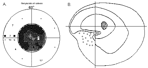
As an example of the differences in the results of different visual fields ar these above recordings of a person who has traumatic defect of the visual pathways. Automated perimetry gives an impression of severely damaged visual field, Goldman perimetry a relatie loss of left lower quadrant, where however responses are present to quickly moving targets (o). this person does not subjectively experience any loss of vision.
- Other visual-sensory functions.
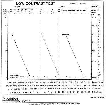
- Contrast sensitivity
ability to see luminance differences on adjacent surfaces (one surface darker than the other). Loss of function at low contrast may be corresponding to the loss of function at high contrast or less or more pronounced than the loss in high contrast vision. Low contrast vision is most important in communication and is also important in other main areas of visual functioning.- Normal vision, visual acuity at 2.5% better than 6/60
(1/10, 0.1, 20/200) (see figure at the end of the document),
- Low vision, visual acuity at 2.5% equal or less than 6/60 (1/10, 0.1, 20/200), better than 6/380 (0.16/10, 0.016, 20/1250),
- severe low vision, visual acuity at 2.5% less than 6/380 (0.16/10, 0.016, 20/1250)
- Low vision, visual acuity at 2.5% equal or less than 6/60 (1/10, 0.1, 20/200), better than 6/380 (0.16/10, 0.016, 20/1250),
-
In case contrast sensitivity is more affected than visual acuity, classification is based on contrast sensitivity loss.
Colour vision
the ability to recognise and match colours. It is screened in normally sighted individuals by using pseudoisochromatic plates and assessed by pigment matching tasks. Classification is always based on assessment. Confusion of colours can be mild, moderate, severe or total.Motion detection and discrimination
the ability to detect that an object is moving and to be able to notice changes in the speed of a moving object. Loss of motion information can be caused by retinal lesions, lesions in the visual pathways or in the cortical functions. Because of the lack of clinical tests, motion detection and discrimination can only be described as normal, subnormal or lacking.Light sensitivity
the ability to perceive light. incl. dark adaptation. Clinically and functionally the most important measurements are measurement of speed of cone adaptation and the level of adaptation reached in half an hour. Adaptation toward lower luminance levels can be delayed, partial or lacking. Photophobia can be mild, moderate or severe.Other specified
incl. vitreous floaters, distortion of image due to changes in optic media or in the retina, entoptic phenomena (= flashes of light, slowly moving glowing scotomas), monocular diplopia due to irregular refractive surfaces. Also ptosis, if it prevents using the eye.Unspecified
- Normal vision, visual acuity at 2.5% better than 6/60
(1/10, 0.1, 20/200) (see figure at the end of the document),
-
The paragraph Abnormal sensation of the eye and adjoining structures does not belong to measurable impairments. These are symptoms that, however, may affect activities.
Other specified
Unspecified
-
Discussion:
There should be guide lines for how the different visual functions are summed, what is the weight of the different components. Above I have stated that contrast sensitivity is a more important variable than visual acuity if there is difference in the degree of loss of these two functions (=Type III loss of contrast sensitivity). One possibility might be to state: The classification is based on the visual acuity values, modified by the effect of defects in other visual functions. Thus for example a person with visual acuity 0.2 at full contrast and 0.063 at 2.5% contrast, nystagmus, ring scotoma that affects mobility and delayed dark adaptation would be classified as having severe low vision.
Comments!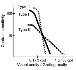
Visual acuity of near vision
Visual acuity at near is defined similarly to visual acuity at distance and
is measured with corresponding line tests using the same optotypes
that are used in the distance visual acuity test.
Groups Severe low vision, Profound low vision and Total blindness are for statistical purposes grouped to one group Persons needing services for the blind.
Discussion:
There should be a statement on what to record when visual acuity at distance is different
from visual acuity at near. One possibility would be to divide the sum of them by two, or, in
order to give more value on near vision functions, add distance visual acuity to 2x the near
vision acuity and divide the sum by three.