[ 1-18 ]|[ 19-37 ]|[ 38-56 ]|[ 57-64 ]
Transdisciplinary Assessment of Vision II


In the assessment of impaired vision, ophthalmologist's work is supported by several co-workers in hospitals or in private offices. However, we also need help from families, vision teachers and therapists, especially to assess young children with brain damage related changes in cognitive vision. The teachers and therapists, on their part, usually appreciate the opportunity to learn from their own problem cases with damage to one or several specific higher visual functions.

We often think on vision as the function of the eyes, but vision is a brain function, a learned function composed of more than thirty specific part functions. Three key features of parallel visual functioning are crystallised in this slide:
- visual information from the eyes to the cortex is transferred through two pathways: the retinocalcarine pathway over the lateral geniculate nucleus and through the tectal pathway bypassing the primary visual cortex and its form analysis,
- in both these pathways there are two different routes with different functions, the magnocellular pathway, marked with M, that carries all motion information and black-and-white information at low contrast levels but no colour information, and the parvocellular pathway, marked with P, that carries all colour information and black-and-white information at high contrast levels,
- from the primary visual cortex visual information flows in two main directions, upwards toward the parietal lobe as the dorsal stream and downwards to the inferotemporal cortex as the ventral stream.
Because of these parallel pathways, the changes in visual functioning can show a great variation and are often difficult to interpret.

Within the dorsal stream are such important visual functions as spatial awareness, orientation in space and eye-hand-coordination, within the ventral stream many recognition and matching functions, colour and motion perception. Use of visual information may be damaged in any of these specific cortical areas independent from other functions.

As an example we may take the length of straight lines: a three year old child can point correctly when asked to point to the longer rectangle. A child, who does not see the difference in the length of the rectangles, may be able to learn to use haptic information to feel the difference and to create the concepts longer - shorter based on haptic information. If he then still does not see the difference between a longer and a shorter rectangle, he has a specific loss of function of the cortical area that is responsible for perception of length of straight lines.

In such a case it is important to ask the child to grasp a rectangle and to watch the child's fingers. If the distance between the fingers is correct well before they touch the sides of the rectangle, the parietal visual functions can "see" the length and convey the information to planning of hand movements although the child does not have subjective visual experience of the size. This is a typical example of parallel use of visual information in the ventral and the dorsal stream.

Efron designed a set of rectangles that have equal surface size. Thus the total amount of light reflected from the surface cannot be used to see the difference in the form. I have redesigned these rectangles to function as a plaything so that we can observe and train children when they compare the rectangles.

A young patient of mine whose hand you see in this picture taught me a nice way of playing with the rectangles. He told me: "If you examine a child who is not as bright as I am, you could build of the rectangles of one colour a stair like this and ask the child to place rectangles of another colour on it."

This play situation is possible even if the child's hands do not function. Of course we cannot then assess eye-hand co-ordination but we can assess his ability to perceive length when he points with his gaze to choose the correct rectangle to be placed on the stair by the therapist.

Similar to seeing size, perception of line orientation is a specific visual function. This picture from a paper by Hubel and Wiesel in 1968 shows variation of activity of a cell when an edge moves within its receptive field in different orientations.

If we redraw the picture and simplify it, we see that one orientation of a line is related to a few spikes being generated in a cell, whereas another orientation of the same line causes much greater activity in the cell. Information from all the orientation sensitive cells, when put together, is the basis for seeing lines of different orientations. This information is also used in both the ventral and the dorsal stream functions.

The LEA Mailbox was designed for assessment of perception of orientation of lines. To drop a card through the slot is fun. You give the card in another orientation than the orientation of the slot and watch whether the child turns the card in correct orientation as soon as the hand starts to move toward the slot. Usually the card is in correct orientation before half of the distance to the slot has been crossed.
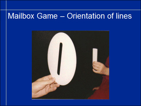
The purely visual task to define the orientation may be possible if the child can turn a rectangle, a rod or his/her hand in the same orientation as the slot without trying to drop it through the slot. If the child finds the task too strange, an adult can pretend not knowing when the object is in correct orientation and asks the child for help by saying "Is this ruler now in the same direction as the slot?"

Orientation of lines may thus be present or absent in higher visual functions either in the ventral or the dorsal stream or both.
Lack of specific visual information in only some higher functions is possible because of the parallel pathways. Sometimes the dorsal stream functions have not been damaged at all while the ventral stream functions are totally or nearly totally lost. Such a child may move without bumping into obstacles, may find his way and grasp objects with hand movements clearly based on visual information, yet he may not be able to recognise forms or name colours. These are the children whom we call "sighted blind". In some functions they use techniques typical to blind and in other tasks they are sighted. They are a challenge to their special educators and classroom teachers.

Among children who have problems in perceiving basic components of pictures are many children with cerebral palsy (CP). I this case there was no difficulty to see the difference between a longer and a shorter rectangle. Drawing a line parallel to another line was also possible, even if the boy did not have his own pen and support for his hand was poor.
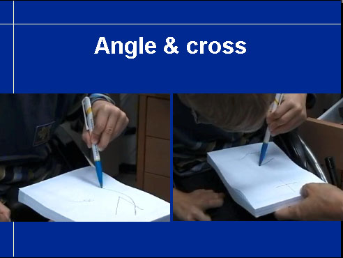
When he should draw an angle it was not possible, also the second time he failed to copy the angle. Drawing a cross was even more difficult. Cross is often one of the earliest successfully copied forms but in this case there is little in common with the cross that I drew for him and the form that he drew.

Here we see all three drawings: rather good parallel line and unsuccessful trials to draw an angle and a cross.

In this video you see his difficulty to fixate his gaze, which is a problem of many children with cerebral palsy and not always diagnosed. Fixation, saccades, the quick eye movement from one fixation to the next, and accommodation are three important motor functions that should be carefully assessed in preschool examinations and at the latest when the child is starting to learn to read.
The slide on the right depicts measurement of accommodation when the boy is asked to look at an accommodative target next to the retinoscope.