Fluorescein and indocyanine angiography
|
|
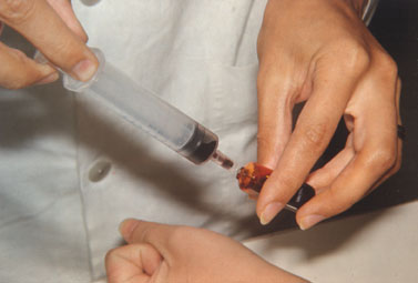
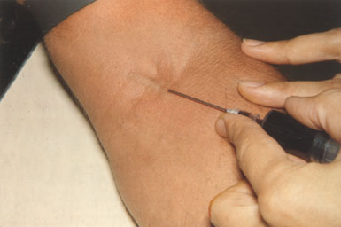
Fluorescein dye is injected in the vein and reaches the eye in a few seconds. When it is circulating in the retinal vessels even the minute capillaries can be visualised, i.e. the blood in them is visible because of the dye. If the structure of the vessels has changed as in diabetes where some capillaries may close, other capillaries may become wider than normally and in some areas the capillary wall may let the dye to enter the surrounding retina, the changes are clearly visible in the angiogram.
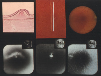 This kind of large composite slides we used at the Wilmer Institute in late 60s when we were learning about the relationships between histopathology and fluoresceinangiographic changes of retinal disorders. In this case the angiogram shows diffusion of the dye through a minute hole i the pigment epithelium into the small detachment of internal retinal layers and the slow spreading of the dye into the small fluid filled space in the retina.
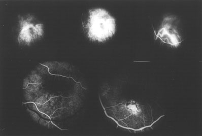 Indocyanine angiography was started in late 60s at the Wilmer Institute and a few years later the first larger clinical series were done at the University of Oulu in Finland. These first angiograms done with both dyes, indocyanine and fluorescein do not have the same resolution as today's angiograms but taught us a lot of choroidal circulation, especially the feeder vessels to the subretinal neovascularisations.