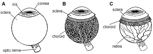
Figure 1 A. When eye muscles and the fat surrounding the eye ball are removed, the white sclera is visible. In the front of the eye its layers are better organized and the wall of the eye becomes the clear cornea. In the back of the eye you see part of the optic nerve that transfers visual information to the brain.
B. When the sclera is removed, we see the choroid, the layer of vessels that bring nutrition to the outer layers of the retina.
C. When the choroid is removed we see the thin, transparent retina with its vessels. Inside the retina there is vitreous that fills the entire cavity of the eye.