Levels of visual pathway lesions
Before the diagnostic observations in the functional assessment can be discussed, a short overview of the visual pathways is necessary to give a common ground to the discussion. During many international courses, visual pathways have been the most difficult chapter. Therefore the simplified pictures on the visual pathways were drawn for the "Vision Testing Manual". These pathways should be drawn by the reader as many times as is needed until their structure is quite clear. Otherwise it is too difficult to understand how a lesion might affect function.
Before attempting a functional assessment or a clinical evaluation of a child's vision we should try to have a clear picture of the anatomy of the lesion(s).

Make photocopies of this drawing (PDF-file) for your training to draw visual pathways.
The following schematic drawing shows the principal components of the visual pathways, as seen from the side of the head.
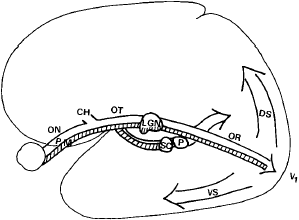 The main, conscious pathway for vision is from the retina via the lateral geniculate nucleus (LGN) to the primary visual cortex (=V1). The subcortical, tectal pathway transfers information to the superior colliculus (SC), and to many nuclei in the brain stem and via pulvinar (PU) to the visual associative functions. From V1 visual information flows upwards as the dorsal stream (DS) and toward the inferotemporal lobe as the ventral stream (VS). Lesions at different locations cause different functional losses. The two main types of nerve fibres in the optic nerve are the parvo- (P) and the magnocellular (M) fibres.
When there is damage to the optic nerve (ON), the change in visual function occurs only in that eye.
When it is close to the chiasm (CH) in the optic tract (OT), it causes loss of visual field on the opposite side. If both optic tracts are damaged, the person has no visual perception but light related diurnal rhythm is still possible, if visual information can get to the pineal body via the small suprachiasmatic nucleus (supra=above, nucleus above the chiasm).
When the lesion is in the optic tract close to the lateral geniculate nucleus, visual information can enter the tectal pathway and via it to cortical functions, apparently only in the dorsal stream (DS) toward parietal visual functions that are related to egocentric and allocentric visual space and visuomotor functions.- When the lesion is in the optic radiation (OR), total damage to the pathway is rare. More often there are small lesions causing a Swiss cheese like loss of fibers with corresponding patchy loss of visual field.
When the lesion is in the visual cortex, it results in loss of part or one half of the contralateral visual field (visual half field on the opposite side). Diffuse metabolic changes may cause loss of certain type of information without permanent field defects.
When the changes are in the secondary or higher visual analysis, they lead to losses of specific functions, not to visual field defects. Damage to the temporal, or ventral, stream (VS) results in agnosias, losses of different recognition functions, whereas loss of function in the parietal, dorsal stream leads to changes in visuomotor and oculomotor functions, and egocentric and allocentric orientation. Visual apraxia is a rare clinical entity caused by changes in this part of visual pathways.
The ventral stream functions are sometimes called "what" functions, and the dorsal stream functions "where" functions. This is an oversimplification because we need recognition of the environment to orient in it although during the movement we do not analyse in detail the structure of the body part moving or the environment. The two main types of vision combine their functions and support them with information from visual memory.
Visual pathway transfers different types of visual information from the retina via different nerve fibers. Parvocellular (P) fibers transfer colour and high contrast black-and-white detail information. Magnocellular fibers (M) transfer motion information and low contrast black-and-white information. The dichotomy is present already at the retinal level, where cone cell and rod cell based vision affect each other.
The presence of parallel visual pathways and functions is typical to the visual system. This feature makes it possible that some visual functions are lost or altered by a lesion and at the same time other visual functions remain normal.
The case history and the current imaging techniques give enough information for a good start. In developing countries, assessment must be based on careful and detailed history taking, general clinical examination, ophthalmologic examination, if available, and on the observations during follow-up.
When we make our observations on children's or adult persons' visual functioning, we should have this variation in size and location of pathway lesions clear in our minds. Pathway lesions are easily forgotten, when the child has a lesion in the eyes. However, for example, many children with ROP have greater perceptual than primary visual problems.
When we list the most important details that we should know about a child we need to pay attention to both cortical and subcortical functions. In the functional evaluation the questions are thus:
- how does vision loss affect this child's communication
are there other disordered functions that also affect communication - how does vision loss affect this child's orientation in space
are there other disordered functions that affect orientation - how does vision loss affect this child's ADL functions
are there other disordered functions that affect ADL functions - how does vision loss affect this child's sustained near vision tasks
are there other disordered functions that affect near vision tasks
Variation in the combinations of the different disorders and in their severity is great. At one extreme are the children whose handling of visual information is so chaotic that they tend not to use it at all. At the other extreme are children with changes in one recognition function, an isolated agnosia, that can be identified by careful history taking and observation but the disorder does not affect the child's functioning other than in that specific task. The variation in the changes in the other sensory and motor functions and memory is equally great. Some children have delays in cognitive development or psychiatric disease complicating the picture.
In some children visual experiences do not seem to be pleasant, they appear not to enjoy looking at things . This phenomenon is known in adult patients with neurological disorders who after circulatory failures in the brain stem sometimes experience a loss of pleasure in looking. Such a loss may last for months. If a similar lesion has occurred in a child who therefore does not experience pleasure when using vision, vision cannot function as the driving force as it does in infants and children with normal brain function. How often we have this problem in children who do not show interest in looking at things, is not possible to guess.
The function of looking at things is a specific function that requires directing attention to the object. If there is a deficit in directing attention to visual information, the child does not pause to look at some specific features, the gaze glides from one object to another. This feature is also difficult to diagnose with certainty. It should not be confused with lack of focused looking caused by insufficient accommodation. Children are not routinely assessed for their accommodation although in many cases it is possible to measure whether there is any change in refraction when an interesting object is moved closer and farther away. Therefore, always, when a child does not seem to look at things or focusing does not seem to be adequate, but the child seems to look through, not at the object, the effect of plus lenses on the child's behaviour needs to be investigated.
Autistic features are often inappropriately mentioned as a feature of children with CVD. They may have true autistic features but in a number of cases the diagnosis is based on misinterpretation of the child's visual behaviour. If a child cannot direct gaze at another person but the gaze wanders on both sides of him/her without eye contact, it may be inappropriately diagnosed as autistic behaviour by someone who is not familiar with the child's problem. Similarly, a child who uses eccentric fixation and seems to look past the person - often at or above the hairline - and not at the person to whom he/she talks, may be incorrectly labeled as "avoiding eye contact". A child who does not accommodate to focus on the other person's face, seems to look through but not at the person and this behaviour may again be incorrectly labeled as an autistic feature.
Before the assessment, we should discuss the behaviour of the child in detail. Children with CVD have several behaviours in common. A particular child may have a few such features, while another may display nearly all of them, depending on which brain functions have been altered. Observations of parents, therapists and teachers can be collected by using a questionnaire covering the following aspects of behaviour:
- Variation of visual functioning is the most common feature
- early development of speech can occur as a compensatory function
- effective use of memory as a compensatory function
- the child prefers talking with an adult to playing in a group of children
- the child dislikes crowded places, and clings to the parents; beaches and swimming halls are worst if recognition of faces is difficult
- the child starts drawing and painting late or never
- colours are used for coding more than by normally sighted children
- there is little interest in TV and comic series
- the child may show signs of spatial interpretation problems , stops at thresholds and shadows
- the child may learn letters and numbers early but may not learn to read except short words
- the child uses siblings and adult persons as helpers when there is a demanding visual task
- the child may become angry when someone moves her/his playthings or clothes even minimally.
Instead of using printed forms that the parents, therapists or teachers would fill in, it is advisable to collect this information during interviews. It would be a great improvement if these questions were asked at strabismus (squint) clinics because so many of these children are treated there without being diagnosed as having the more important deviations of brain functions.
The compensatory techniques used in pre-school activities are not possible in school situations. Therefore school often becomes a great disappointment and misery to a child who was an eager learner in pre-school.
Previous Chapter 
|
 Next Chapter Next Chapter
|