[ 1-19 ]|[ 20-39 ]|[ 40-59 ]|[ 60-79 ]|[ 80-100 ]
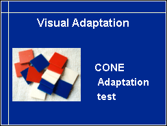
Visual adaptation to changes in luminance level is affected in several diffuse retinal degenerations. The speed of cone cell adaptation may become longer years before the child has real night blindness. The test takes almost no time and costs very little so it is a perfect screening tool for retinitis pigmentosa among hearing impaired children. It is also a good test in assessment of all visually impaired children who usually do not like the long clinical tests that measure also rod cell adaptation up to half an hour.
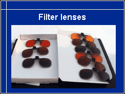
There is one more question that we need to consider during this second lecture. It is filter lenses. Photofobia is a common problem and needs to be treated. In this slide we see a collection of filter lenses that filter short wavelength light with a cutting point at 511, 527 or 550 nm. It is possible to have another filter added to prevent polarised light to get through. Polarised light is light reflected from water and metal surfaces. These filters are manufactured by Multilens in Sweden. The oldest filter lenses of this type are manufactured by Corning and are photochromatic, i.e. they become darker in sunlight.
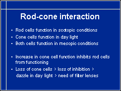
Photofobia that is treated with filter lenses is caused by loss of cone cells in the central retina. Normally cone cells inhibit the rod cells from functioning in daylight. However, if the number of cone cells relative to the number of rod cells becomes too small, then cone cells cannot inhibit the rod cells. The very sensitive rod cells are overexcited in daylight, the person becomes dazzled. The rod cells absorb light most effectively in bluegreen part of the spectrum. When this part is filtered away with the filter lenses, the luminance level is good for the rod cells and at the same time cone cells get enough light to function, so the image looks brighter and calm.
Photofobia can also be caused by cloudy cornea, lens or vitreous that cause glare like dirty windshield does. In these cases filter lenses may or may not be experienced beneficial. However, many persons with cloudy media like yellow filters in their reading lenses to reduce scatter of blue light.
Allergic keratoconjunctivitis causes photofobia that is best treated with regular sunglasses and a broad brim in the hat and proper medication to reduce the symptoms. Filter lenses would increase photofobia in this case because they increase subjectively experienced brightness.

|
|
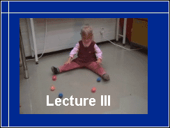
We have two important functions to cover before we can move to the so called cognitive visual dysfunctions or CVI. One is motion perception, the other is visual field. In this video sequence we can observe both motion perception and visual field. First I roll one ball and observe how well the child can follow the movement - there does not seem to be any difficulty. Then several balls are rolled several times while I observe whether the child responds on both sides of her visual field - she notices balls on both sides, reaches for them in similar ways and is even able to copy the way several balls are rolled at once. Conclusion, normal perception of balls rolling, symmetric visual field, good eye-hand-coordination, good copying of complex motor tasks with hands. (See the video.)
Visual field loss may result from lesions in different parts of the visual pathways: retina, optic nerve, optic tract, optic radiation or primary visual cortex, V1. Lesions in associative visual cortices do not cause loss of visual field but loss of specific higher visual functions.
The facts that we should know about visual field are 1.its size, 2. whether there are scotomas, areas of poor function within the visual field, 3. whether the image in the central visual field is normal or distorted, and 4. whether the child experiences any light phenomena that are not part of the image but occur floating in front of it. Features 3 and 4 most children can discuss first at the age of 8-9 years. However, a child may ask "why there are fireflies during winter" in areas where there are fireflies during summer. Then we know that the young child is seeing light phenomena that are caused by abnormal functions in the retina.

Visual field can be measured with many techniques. The technique that is possible everywhere in the world is finger perimetry or confrontation perimetry. You bring your hands behind the ear level of the child, place your right hand behind the upper left visual field of the child and your left hand behind the lower left visual field of the child, ask the child to look at your nose and move your hands forward while moving your finger as quickly as you can. When the child sees the moving fingers, he points with his hand in the direction where he sees the movement. Next you measure the limits of the two other quadrants of the visual field in the same way. (See the video.)

Clinical tests depict visual field in different ways. Perimetric tests have been developed to detect glaucoma early, not to assess functional vision. For example, retinitis pigmentosa causes in young children some loss of function in "pericentral" visual field, in area around the central visual field. When such a change is measured using automated perimetry (on the left in this slide) there seems to be large non-functioning area around the central field, a ringscotoma. However, when the same visual field was measured less than an hour later using Goldmann perimetry, our 'gold standard', there is no scotomatous area present. - We will soon see that also Goldmann visual field may not correctly depict functional visual field.
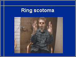
Older children can describe their visual field and demonstrate like this boy in the video that in room light he sees finger movements clearly in the far periphery, then the movement become less distinct in midperiphery and again is clear in central vision. There is relative scotoma to motion perception, although Goldman visual fields showed "absolute ring scotoma". Goldmann visual fields are measured at 10 candelas per square meter, which is a very low luminance level. In brighter light scotomas may shrink notably, even disappear. (See the video.)
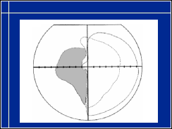
Loss of one half of visual field is common in older adult people but occurs also in brain damaged children. If the lesion is way back in the visual pathways, the tectal pathway may remain intact and transfers information to higher visual functions. If no information comes from the optic radiation to the primary visual cortex, then this cortical area rearranges its connections and may use visual information in ways that we cannot examine with our present techniques. If a child has an accident in school age, it is wise to start stimulation and training soon after the accident.
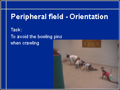
We can create several play situations and games to observe function in the peripheral parts of the visual field. Here, for example, children crawl avoiding the bowling pins. The bowling pins have nearly the same colour as the floor and therefore are ideal in assessment of low contrast vision in the periphery. The youngest child, who seemed to have limited field, does not pay attention to the pins but the therapist needs to help her in the task. Situations like this function as tests and at the same time training to scan the surroundings carefully.
Loss of peripheral visual field can be compensated using careful scanning technique when walking. In physical education and similar situations where unexpected objects may appear in the peripheral visual field the structure of visual field needs to be known by the teacher in charge of the activity. It might be necessary to report it to the insurance company of the school in some countries.
Structure of central vision should be known for all sustained near vision tasks.
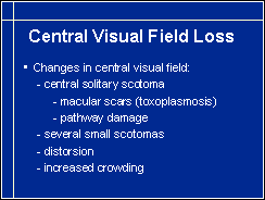
Changes in central visual field affect communication and sustained near vision tasks, like reading and similar demanding near activities. Lesions may be one larger central scotoma or several small scotomas, distortion of the image or increased crowding (= single symbol acuity much better than acuity measured with crowed symbols). Large central scotoma shifts fixation to some better functioning area of the nearby field and the child seems to look past the person whom he looks at. Since people often react negatively to unusual eye contact, this cause for deviation from normal should be explained to all who are in contact with the child.
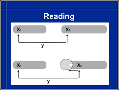
If a small scotoma happens to be on the right side of the point of fixation so that it falls on the next word to be read, it "eats up" a few or only one letter and may disturb planning of saccads when it covers the beginning of the next word. This may lead to errors in reading but the errors do not follow any typical pattern of reading difficulties.
If a child makes atypical reading errors, it is wise to use larger texts of several font sizes. If the errors happen at the same physical distance from the previous fixation even when the text is larger and disappear when the text is large enough, then you know that the child does not have a usual reading problem but is disturbed by a hole in his central visual field. The solution is either to use large enough texts or experiment with the child to find a slightly oblique orientation of the text where the scotoma does not disturb.
As we have seen, visual field needs to be known in detail, not only its size but also its structure in midperiphery and in central vision.
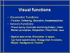
There are some 30 higher, complex visual functions called cognitive visual functions. This name is somewhat misleading, because the visual functions that we have just discussed are also cortical functions. Perception of forms, colours and motion, as well as integrating the different parts of a scene to an image, are all cognitive functions. Cognitive visual functions usually mean higher visual functions like recognition of faces, perception of expressions, perception of objects against a background, spatial orientation, eye-hand coordination etc. Visual functions in the early part of processing visual information like perception of length of straight lines or their orientation are usually not assessed in neuropsychology although they are well known functions that can be lost without a major vision loss.
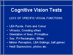
Several higher visual functions can be assessed at school using simple test situations. Similar to the use of the Colour Vision Game to experience which colours are confused by the child, use of these tests at school makes it much easier to understand that it is not an intellectual but a vision problem when the child cannot complete a task.
Some of these tests we have seen during the previous two sessions, like the LEA Puzzle, visual acuity tests, Grating Acuity tests, and ball games, four other tests will now be introduced.