6. Visual Fields

Visual field is the space in front of us when we are looking straight forward without moving our eyes. When we measure the size, the outer edges, of a visual field, we bring the stimulus from behind and watch when the infant/child notices the movement in the peripheral visual field. When we test young infants, it is often difficult to balance attention between central and peripheral stimuli. The infant may be so interested in the face in front of him/her that peripheral information is not noticed at all. Sometimes it is easier to have the infant in a baby sitter in front of a mirror for a while and observe the behaviour when bringing the toys from behind.

Types of measurement techniques vary depending on the communication and developmental level. The measurement technique described in the previous slide is called confrontation. The stimulus can be a toy, small light source or moving fingers. When the child grows, Goldmann perimetry is often possible at the age of four and half to five years.
Automated perimetry is often difficult to perform and does not correctly depict functional vision in retinal degenerations. Eccentric fixation recorder and Macular Mapping tests have been developed for thorough investigation of reading field and become important when the child starts reading.
Flicker perimetry and measurement of discrimination of movement are still experimental methods for assessment of function in the magnocellular pathway. Motion perception can be assessed with figures in motion. Although there are several tests for measurement of the size and structure of visual field, observation during play always gives valuable information.

Visual field loss is due to changes in the retina or in the long visual pathways. It develops slowly in many inherited retinal disorders and thus the child does not notice the gradual change. In such a situation perimetric recordings are important. The size of the visual field does not change early but function is lost in 'midperiphery', not in the middle or at the periphery but half way between. When the sensitivity in midperiphery has decreased, isopter I/4 is shrunken. Normally it is slightly larger than the III/4 isopter is in this case. There are also some hatched areas where the stimulus has sometimes been seen, sometimes not.

As an example of how differently the Goldmann perimetry and an automated perimetry depict visual field in RP are these two perimetric recordings measured only an hour apart. In the automated perimetry there is deep ring scotoma with no response to the brightest test lights, whereas Goldmann perimetry shows a diffuse decrease in sensitivity leading to constriction of the isopters but no ring scotoma. The boy had been tested elsewhere with automated perimetry and had been told not to ride his bike because of tunnel vision. This error seems to be quite common in hospitals and offices where technicians measure visual fields and automated perimetry is the standard test.

Even the Goldmann perimetry does not depict the function of visual field in all cases. In this case there is damage to the visual pathways just in front of the primary visual cortex. In MRI it is visible as a black area on the right side. On the left side in the corresponding site there are the fibres of the visual pathway visible. In Goldmann perimetry there is left loss of half field on the left side with sparing of the macular field. However, the patient saw movement on the left side of the visual field and experienced 'glow' when the largest, brightest stimulus was moved very fast back and forth.

When the left side of the visual field was measured using flicker perimetry, lower than normal but measurable function was documented. After training of the 'blind' visual field for months symmetric flicker sensitivities could be measured. This means that rehabilitation to improve vision is possible in such brain lesions where parts of the pathway remain functioning. In this case it looks as if the tectal visual pathway was spared.

When brain functions were recorded with MEG during stimulation with flickering target, activation was recorded on the normal left side of the brain, both in the primary visual cortex and in temporal cortex. When stimulation was in the left visual field, there was no recordable activation in the primary visual cortex but a delayed response was measurable in the temporal cortex (Vanni et al. 2001)

When the 'blind' area was stimulated with flickering letters, recognition of letters reappeared in the previously poorly functioning area. Where recognition of letters occurs in this case, is not possible to investigate but it gives us a totally new view of possibilities to train, rehabilitate visual functions.

Since improvement of function can occur in an adult person several years after the trauma, it is likely to happen in children whose brain has much greater plasticity than the adult brain.

Although we can measure several functions of the visual field, there are numerous phenomena that are not measurable. When retinal degeneration has destroyed part of the complex connections within the different layers of the retina, there are disturbances in the image quality. This animation tries to depict the 'moving net in front of the image' and the 'sausages' that disturb many persons with retinitis pigmentosa. The speed of the movement varies. Some persons describe 'arrows' moving across the central visual field rather a net.

The 'sausages' are described also as 'bananas'. They appear one by one at a certain point at the edge of the ring scotoma, move around along the edge of the ring scotoma and may be followed by one or more similar lights that all move with the same speed. After one or several circles the lights disappear into the ring scotoma again one by one. All persons who see these light phenomena describe them moving clockwise.

Disturbing light phenomena usually appear after school age but children may see small lights resembling the lights in the automated perimeters before school age. In countries where there are fire flies these children surprise their families by telling that they see fire flies also during winter when there are no fire flies. Sometimes the child may ask why other people are not watching the nice little sparks that appear in twilight.
When the ring scotoma develops, it is subjectively not visible but small things start to disappear on the desk at school and after a while 'reappear' as if somebody was playing games with the child with RP.
Deaf children notice that their participation in group discussions becomes difficult because the important beginning of sentences are missed. They do not notice when somebody starts signing if the person happens to be within the ring scotoma. (In sign language the doer(s) and the object are defined in a certain location of signing and after that the verb signs move between these locations without repeatedly defining the doer or the object. So if the beginning is not seen, the later part of signing may be misunderstood.) When the central visual field becomes a narrow tunnel, the teenager moves backwards to see larger signs, which often is misinterpreted as a sign of failing interest in communication.

In summary, we can list the types of peripheral visual field losses as
1. Hemianopia and quadrantanopia, loss of a half or a quadrant of the visual field.

2. Bitemporal field loss, of the side part of visual field in both eyes.

3. Constriction of the visual field, which may occur because of atrophy or hypoplasia of the optic nerve or ROP, retinopathy of prematurity

4. Altitudinal, which means loss of lower or upper visual field. Loss of the lower part is more common and results in a typical head posture, because the child must bend the head down to see the ground before his/her feet.

5. Loss of upper part of the visual field is often caused by colobomas of both eyes. If the field losses are not symmetric and the functioning part of one field partially compensates the nonfunctioing part of the other field, the area of loss of visual field may be small and thus no functional loss exists. Visual fields need to be assessed as a sum of the two fields if both eyes are used simultaneously. If not, then visual field changes its character whenever the child shifts fixation from the one eye to the other.

6. Visual field defects are sometimes confused with attentional difficulties. If a child cannot direct his/her attention on a certain area in subjective space (s)he seems to have field loss. This is common in young infants with hemiplegia who may have also complete neglect of the hemiplegic side of their body. In these cases it is important to guide the infant's attention from the better functioning side to the less functioning side by using touch and sound. In some children 'hemianopia' disappears or becomes much smaller before the child is two years old. Then we know that there has been enough function to create neuron networks so strong that the visual field can be used for orientation in space and even for demanding near vision tasks.

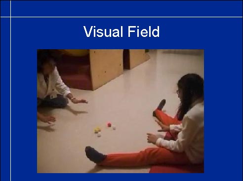
Video
7. Since measurement of visual field is difficult before school age, we need to create play situations where we can observe both central and peripheral visual field. First we have to know that the child can perceive moving objects, has motion perception. Rolling first one and then several balls on the floor reveals whether the child's gaze follows the movement of the ball(s) or makes a quick saccad when the ball(s) stop moving. If the child seems to be able to see the moving balls, several balls are rolled at once. When this is repeated several times it is possible to observe whether the child respond symmetrically, i.e. the two halffields function symmetrically or one of them dominates.
Video
Also older children may be assessed with this simple ball game. Rolling balls on the floor is a better technique than throwing balls to a standing child because motor functions may require so much of brain capacity that catching balls in the air is not possible. How well a child can catch a ball, is often more affected by the motor problems than by the child's capability to visually orient in space and see details in it.

Ball games are useful in investigating children's responses to transient visual information, to observe the following movements of the eyes, whether a child has symmetric responses in both sides of the visual field and how quick and accurate the saccadic movements are.

Video
Peripheral vision can and need to be observed in play situations. In this case we had a suspicion that the youngest of these visually impaired children had restricted visual field. When the children were instructed to crawl 'like hunting indians' without touching the bowling pins, the older children did so but the youngest was not aware of the presence of the pins before instructed to scan the surroundings. Such a situation makes the child aware of the need to develop efficient scanning techniques to compensate for the small size of the visual field. Note that the pins have nearly the same colour as the floor, which makes them difficult to notice if contrast sensitivity is low.

Video
Training cane is a good tool to make the child aware of variations in the surface quality that the child is unable to see. When the cane conveys a difference in the surface, the child bends down to find out what the cause is and learns more about the structures and details of the environment.

Video
Climbing a stone of a few meters is exciting to a young child who has never before played in a forest. We can observe the child's capability to orient in a new space and to move in it.

Video
When a person has had brain damage, in this case due to operation of a brain tumour, visual field may be normal in the clinical measurements but orientation in space, both egocentric and allocentric space may be disturbed. We see here major difficulties in turning the puzzle pieces with their coloured side up and placing them in their cut-outs - a task that an 18 month old infant usually masters. Training requires a lot of patience and creativity to help the youngster to develop compensatory skills in eye-hand coordination.

Macular Mapping is the first test specifically designed for assessment of abnormal reading field at the Smith-Kettlewell Institute by Manfred Mackeben. Letters of different size and contrast are shown for varying stimulation times within 16 degrees of the area around fixation point. If a letter is not seen, the corresponding point in the recording is black. If the letter has been noticed, but perceived wrongly, the point is grey. If the letter is seen correctly the circle is white. This way discrimination of letters is documented in different parts of the preferred retinal locus for fixation. Reading technique can then be trained using the best functioning island of vision.

In this case we knew that visual acuity was low. Therefore presentation time is long and the size of the letters four times larger than the standard letter size. There is an island of vision four degrees to the right and six degrees to the left of fixation and similar diameter also vertically.

When the exposure time is shorter and the letter size only slightly larger than the standard size, the functional visual field decreases to two degrees to the right and four degrees to the left. This explains reading difficulties: the small island of vision with low recognition acuity causes problems in planning saccades and in reading longer words. If contrast is increased by using a CCTV smaller texts can be read and thus a greater number of letters is visible at one time.

When a person has a central scotoma that covers foveal area, the person must use eccentric fixation in reading. If loss of visual field is patchy there may be several islands of vision that are preferred in different visual tasks. The island that is used when measuring visual acuity may be too small or distorted for reading text and a more peripheral island with lower visual acuity but better image quality may function better. Shifts in fixation can be documented by using SLO, scanning laser ophthalmoscope but it is so expensive that only few places can afford to buy it. Therefore I have designed the Poor man's SLO.

Poor man's SLO, the Extrafoveal Fixation-recorder, is a derivate of Amsler Chart. In the middle of the chart there is a hole, where words or pictures of different size can be placed. When the person is fixating on the word the test is slowly moved closer to and farther from the person who at the same time observes whether the dot, cross and diamond disappear and reappear. If they remain visible all the time the blind spot has been shifted from its usual location. It can be mapped the same way as in Goldmann perimetry. When the location has been found, the text is changed to a smaller or a larger text or the luminance level is changed. The location of the blind spot is mapped again. If it is now at a different location than during the first measurement, the fixation has changed as much as the location of the blind spot has changed. The distance between the eye and the test needs to be kept unchanged.

Direct observation of fixation and saccadic movements often is enough to reveal the difficulties in reading. In this case the girl seems to look at her finger in front of her eye but she is fixating a word in the middle of the text. The only attached area of the retina is in the nasal side of the optic disc. - When the text is written close to the edge of paper, the eye movements and fixation are easy to observe.

Reading is composed of fixations (X1 and X2)and quick saccades to the next word or to another part of a longer word. While a beginning reader spells the first word letter by letter, the distance to the beginning of the next word changes constantly, yet the visual system is able to calculate the length of the next saccade when the child has read the word aloud or silently, at which time fixation may move backwards to the centre of the word. The visual 'map' information is passed to the motor functions and a motor plan is made. Before it can be executed, a command to detach gaze from the present fixation must be given. After that the saccade brings the gaze to the beginning of the next word (adult persons fixate more toward the middle of the word), it is fixated there and then moved with small saccades along the word if the child still reads each letter separately or the word is recognized during one or two fixations.
If there is a scotoma on the right side of the point of fixation, it may now and then interfere with reading if it happens to fall on the beginning of the next word. In that situation the beginning of the word is not visible, the saccade is planned to the first visible letter and a correcting saccade backwards is needed to begin from the beginning of the word. - If a child recognizes words without spelling, a scotoma on a word makes one or several letters to disappear, which leads to 'misspelling'.

When a child makes errors in reading age appropriate texts, the presence of disturbing scotomas and distortions can be revealed by varying the size of texts. If the errors disappear when the size increases then the errors are related to poor image quality. This simple test should be done in each case before special tutoring in reading is started.
Other important observations on the quality of the reading field are 1. the size of the reading field, i.e. how many letters are visible at a glance; 2. whether there are distortions of the text (this requires an experienced reader to be able to analyse variations in the forms of the characters): 3. size of communication field, reading signs, when the child is hearing impaired.

Central solitary scotomas cause eccentric fixation, use of a preferred extrafoveal locus, which may not be the best area for all sustained near vision tasks.

The causes of solitary central scotomas vary. Macular scars caused by infections or large macular hemorrhages are common in the younger groups of children. At school age, loss of macular function may be caused by an inherited disorder or trauma and thus early intervention is started in kindergarten or school age.

Central scotoma may be caused by damage to the visual pathways or primary cortex.

Sometimes there are several small scotomas that distort the image even if visual acuity may be good.

Distorsion may be caused by folds in the retina or minute changes in the foveal cones. If they are tilted after foveal oedema, image quality is affected.

Increased crowding is a feature of central visual field that should always lead to thorough investigation of the structure of the posterior visual pathways and primary visual cortex. Increased crowding means a greater need of magnification than would be expected based on the routine line acuity.

In summary, the assessment techniques used in measurement of visual field size are confrontation tests and the Goldmann perimetry.

Other qualities of the visual field can be assessed by using flicker perimetry, Macular Mapping or Extrafoveal Fixation Recorder, and the quality of the reading field by measurement of visual acuity with single symbols, line test and crowded tests.

However, disturbing lights in the visual field, trembling of the image and other varying changes in the image need to be described by the person because they cannot be measured with any tests.