
To have a glimpse of magnocellular information we can look at these moving gratings or

...a flickering spot of light. Both represent transient, rapidly changing visual information.

About 80% of the fibres in the optic nerve are parvocellular fibres. They transfer information from small retinal ganglion cells to small cells in the lateral geniculate nucleus and from there to the primary visual cortex. These fibres are thin and have therefore lower speed of transfer than the magnocellular fibres. They carry all colour information and are effective in transferring high contrast black-and white information.

The next two slides depict some differences between parvocellular and magnocellular information. You see that there is a small grey dot in the upper right hand corner and a stop sign in the lower left hand corner. If you look at the stop sign and slowly count to five, the grey spot has disappeared from the image you see although it is still there.

If you keep on looking on the stop sign, you notice that there now appears a flickering spot that does not disappear like the non-flickering grey spot. On the contrary, it makes the non-flickering spot visible now and then. Visual system is more effective, more alert to transient information, information in motion, than to non-moving visual information.

This picture is from Margaret Livingstone's and David Hubel's publication in 1988 depicting the parallel pathways for magnocellular and parvocellular information via the lateral geniculate nucleus to the primary visual cortex where they enter in different cortical layers. Although this information has been known for more than twenty years, it has first recently become a part of clinical examinations. These different parts of the pathway are important to consider, because changes in magnocellular pathway may affect only low contrast vision, contrast sensitivity. If contrast sensitivity is not measured, the changes in the magnocellular pathway are not recorded and the person may be told to have as good vision as before or may be taken as neurotic. On the other hand, small changes in the middle of the visual field may cause loss of visual acuity without major loss of contrast sensitivity.
Within the primary visual cortex there are cell groups(the so called 'blobs') that are activated by colour information, other groups of cells that are activated by form information (in the interblob areas) and yet a third type of cell groups that are activated by visual information in motion. From the primary visual cortex information flows in several specific areas of the visual cortices after which its different components are brought together and fused in still higher visual functions and an image is perceived.

This simplified picture of the previous drawing shows the three target areas of visual information in the primary visual cortex, the "blobs" of cells for colour, interblob areas for form information and the layer for motion related information.

From the V1 area, visual information flows in all directions. The two main directions of the flow are called "dorsal" and "ventral" stream. Dorsal stream upwards to the parietal lobe and from there to the frontal lobe, and ventral stream down and forwards to the inferotemporal lobe, the lower part of the temporal lobe.

In these two main streams of information, magnocellular information dominates in the dorsal or parietal stream and parvocellular information dominates in the ventral or inferotemporal stream. There is an effective "communication" between the different visual cortical areas and a mixture of the different types of visual information so that when finally the image is formed, its form, colour and movement are seen fused to a clear picture.

If the connections between the different levels of higher visual functions are drawn as we see the structures from the side of the head, it looks like this. Again, I would like to stress the effective connections between the specific cortical areas that are in charge of more than thirty visual subfunctions. In this picture there are only a few lines depicting the myriads of connections between the different cell groups.

On the surface of the brain, the visual cortical areas are both in the occipital, in the temporal and in the parietal lobe. There are also important visuomotor areas in the frontal cortex, the so called frontal eye fields.
With these slides on the structure of the visual pathways I wish to create an awareness on the multitude of functions that we need to observe and measure, not only visual acuity. Since brain damage has become a common cause of visual impairment, we need to develop testing and observation of higher visual functions, cognitive vision or visual perception. Visual acuity is one of many visual recognition functions.

In the two main streams of visual information, eye-hand-coordination and spatial awareness are functions in the dorsal stream, whereas the numerous recognition functions are in the ventral stream area.

At all levels of transfer and processing in the visual pathways there are parallel functions. This is important to remember when we assess visual performance of an infant or a child so that we assess each of the parallel functions at every level. In the sensory retina we have cone cells and rod cells, the cells for day vision and night vision. From the retina the information is transferred through the parvocellular and the magnocellular pathway in the retinocalcarine and the tectal pathway to the brain cortex where it is spread via the dorsal and the ventral stream to cortical functions. The cortically "analysed" information is used also in the subcortical functions.

When we start an assessment, we first collect all clinical information available. Ophthalmologists, optometrists, low vision therapists, other therapists, and paediatric neurologists report to the child's paediatrician, who functions as the coordinator of the services. In the ophthalmologists' reports the most important findings are the description of the anatomic changes or malformations, refraction and - if glasses have been prescribed - their values and at which distance do they make the image clearest.
If the lenses are under- or overcorrected, a description is needed of why it was done and at which distance the image is clearest with that correction. This is especially important if an infant is aphakic, has no lens in the eye, because without the lens, the eye cannot focus the image the same way as a normal eye. Infants and children, who do not accommodate, need to have near correction, reading lenses. Reading lenses blur the image at distance, which needs to be known by all persons who take care of the infant.
Few visually impaired children are fully binocular. Many children have strabismus and those children whose eyes are ortotropic, "straight" eyes, may have unequal quality of the central image in the left and the right eye and therefore do not develop binocularity. If an infant has developed binocular vision before vision is impaired, there may be binocularity of varying degrees.

In the assessment we need to consider the use of vision in all areas of development. During the preschool years these functions are the most important ones to consider: communication and interaction, motor functions, spatial concepts and orientation in space, object permanence and language. We need to ask ourselves:
- how is vision affecting each of these functions today
- how will it affect development of vision to its next milestone
- how will it affect development of each of these functions
- what shall we do to enhance use of vision and to develop compensatory techniques of learning in areas where visual information is insufficient.
Special education starts at age zero, not at school age.

This picture from the book by Anne Nafstad and Inger Roedbroe depicts early interaction as a strongly visual function between mother and child.
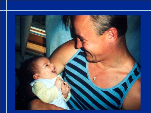
Visual communication is equally important between father and infant.

(Video)
At the age of eight weeks an infant can interact with both parents at the same time and uses vocalisations with clear variations in intonation. (Pause to listen to the infant.) There is no question about the importance of vision in early interaction between this infant and her parents.

At this age we can roughly assess the quality of image by measuring grating acuity and contrast sensitivity.