Transdisciplinary Assessment of Vision I

(Video demonstrating collaboration between a special education teacher and an ophthalmologist to assess a young child with Down Syndrome.)
This video sequence depicts evaluation of visual acuity of a young child as a collaborative effort of a kindergarten teacher and an ophthalmologist. The child accepts her familiar teacher as a partner in the diagnostic play situation. The ophthalmologist observes the responses of the child. Note that at the end of the sequence the child signs "good" as a sign of how proud she is of her performance in the game. This video sequence was taken to document the assessment of this child and is one of the numerous video sequences that have been analysed by the multidisciplinary teams in European countries where we are developing the transdisciplinary assessment techniques.

Assessment of vision for early intervention and educational purposes is a transdisciplinary work that requires medical, psychological and educational expertise. When examination of visual functions is difficult, teachers and therapists are needed to train the child and often also to test the child, either alone or together with the ophthalmologist.

|

|
| Slide 5. | |

|
|
Because visually impaired children are now integrated in local day care services and schools, a great number of doctors, therapists, teachers and psychologists need further education in assessment of vision and in special education. Each child should have a local team of people trained in early intervention and special education of visually impaired children so that a true inclusion would be possible. The new International Classification of Functioning, Disability and Health (ICF) stresses assessment of functioning, activities and participation. This type of assessment requires good cooperation between medical and educational experts.
Increasing number of multihandicapped infants and children and an increase in brain damage related loss of higher visual functions have changed the content of assessment and intervention. The complicated picture of multihandicap is with us to stay. Thus we need to learn about the varying picture of visual impairment and disability and their effect on activities of these children.
At least 70% of visually impaired children have another impairment or chronic illness. Many children have more than four different impairments. In some countries and in some states of the US, a child who is classified as multihandicapped, is not always even examined for sensory impairments because they seen of less importance than the ‘primary’ impairment of multihandicap. This, of course, is not logical, because the degree of multihandicap would be less if sensory problems were taken care of. For example, in the large group of children with Down children, both visual and auditory impairment are much more common than in general population of the same age. Also children with more visible motor problems are likely to have ‘hidden’ visual problems. These problems are ‘hidden’ because children with motor problems are not thoroughly examined for their well know typical losses of visual functioning.

|
|
| Slide 8. | |

|
|
In the assessment of low vision we have three stages or three goals:
- first to understand the quality of the image that the child uses,
- second to understand how the child uses the images in cognitive visual functions
- third, how changes in image quality and/or higher visual functions affect the child's development and learning. Understanding the type of visual functioning is essential in communication and in planning the use of learning strategies in special education. Thorough assessment is especially important if the child will be in integrated education where the classroom teacher has little time for the child and little basic knowledge in special education.

|

|
| Slide 11. | |

|
|
To better understand the structure of visual images, we need to discuss their components. Visual images contain three types of information: forms, colours and motion. We also measure visual field, visual adaptation and oculomotor functions.
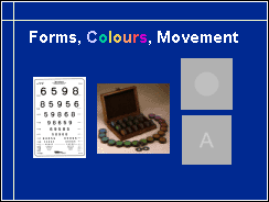
Therefore assessment also needs to cover each of these three types of visual information. We use measurement of visual acuity as a measure of form perception, quantitative colour vision tests to assess colour perception but rarely if ever assess motion perception. It is, however, an important component of visual information because nearly all visual information is in motion. Either the target or the observer moves. If they both are stable, the eyes of the observer move to scan the environment and move with short saccadic movements during reading.
Our most common clinical measurement, measurement of visual acuity should use tests with logarithmic lay-out (as in the test in this slide) both at distance and at near (ICO recommendation 1984, WHO recommendation 2003, link to article on front page). However, tests with non-standardised structure are used in most countries and the visual acuity written in the documents does not say at which distance the measurement was made and whether the test was a line test or a single optotype test. This is as correct as would be documentation of an child’s height with two numbers, not stating whether they refer to centimetres or inches.

In the assessment we use both clinical tests and observations on the infant's/child's behaviours in usual daily activities. Use of vision tests is not restricted to the offices of eye doctors. On the contrary, testing as a part of play situations or classroom work often allows measurement of functions months or even years before it is possible in a doctor's office. After the successful measurements, the interpretation of the results requires consultation by an ophthalmologist.
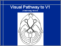
To have a common ground in discussions on visual functions, let us look at a few pictures of the visual pathways. This picture is well known to all of us. There is, however, one detail that might be new: the visual pathway from the eyes to the primary visual cortex is a two way street in its posterior part. There is information flow from the primary visual cortex to the lateral geniculate nucleus (LGN) and the superior colliculus (SC).
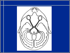
Actually, the pathway between the lateral geniculate nucleus (LGN) and the visual cortecies has far more nerve fibres from the visual cortecies to the LGN than from the LGN to the V1. Thus ongoing processes in the visual cortecies affect the flow of information from the LGN. Only visual information that is ‘needed’ in the ongoing processing is given entrance. This inhibitory function changes the incoming information so that we see what we expect to see.
Visual information from both eyes is transferred to the visual cortex in parallel nerve fibres and fused first in the primary visual cortex. Fusion occurs, if the child uses both eyes and has learned to use them together, binocularly.
For more information go to Visual Pathways.
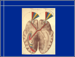
Visual information from the left halves of the retinas of both eyes is transferred to the left half of the brain, the left hemisphere. From the left eye the fibres go directly, from the right eye the cross the midline in the chiasm. From the right halves of the retinas information is transferred to the right hemisphere.
This picture also depicts that macular vision, the central 10-13 degrees, (marked with red in the slide) uses nearly half of the primary visual cortex. The central part of visual image is overrepresented in brain functions. The central 12-13 degrees use nearly one half of all the cells in the primary visual cortex.
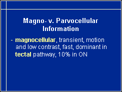
In the optic pathway there are different types of nerve fibres. The two large groups of nerve fibres are the parvocellular and the magnocellular fibres. The magnocellular fibres transfer information from the large retinal ganglion cells to large cells in the lateral geniculate nucleus. These fibres are thick and transfer information with relatively high speed. This pathway carries all motion information and is efficient in transferring low contrast black and white information. It does not carry colour information. In the optic nerve approximately 10% of the fibres are these thick fibres.
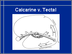
Magnocellular pathway is dominant in the tectal part of visual pathways that leaves the main retino-calcarine pathway before the lateral geniculate nucleus, enters superior colliculus, many subcortical nuclei and via pulvinar into the cortical visual functions bypassing the primary visual cortex, V1.