DIFFERENT VISUAL FUNCTIONS
Vision is composed of many simultaneous functions. If vision is normal, seeing is so effortless that we do not notice the different visual functions.
The different components of the visual image are:
- forms
- colours
- movement.
We see both during the day light and during very dim light. In day light, photopic vision, we perceive colours because of function of the cone cells; in very dim light, scotopic vision, we see only shades of gray, since rod cells respond only to luminance differences. In twilight, when both rod and cone cells function, we have mesopic vision.
Vision is measured with many different tests. In the following chapters we will discuss such tests and measurements that are often done in the doctors' offices and in the hospital clinics: visual acuity, visual field, contrast sensitivity, colour vision, visual adaptation to different luminance levels, binocular vision and stereoscopic vision.
Visual acuity
Visual acuity is measured with visual acuity charts at distance and at near. The test measures what is the smallest letter, number or picture size that the patient still sees correctly. Visual acuity is good only in the very middle of the retina (Fig.20).
Fig.20. In the middle of the retina there is a small pit, the fovea, with which we see sharply. Only a few millimeters from the fovea (arrows) the visual acuity is 20/200 (6/60 or 0.1) even in a normal person. |

|
Visual field
When a person with normal vision looks straight forward without moving the eyes, he sees also on both sides. The area visible at once, without moving the eyes, is called visual field. Nerve fibres from both eyes are divided so that fibers from the right half of both eyes reach the right half of the brain and fibers from the left half of both eyes the left half of the brain (Fig.21).

|
Fig.21. Visual pathways from the eyes to the visual cortex. Note that there are also connections to the central parts of the brain. Note also that in the optic radiation, the pathway from the LGN (lateral geniculate nucleus) to the primary visual cortex there are marked arrows in the direction from the primary visual cortex to the LGN. Actually, there are some ten times more fibres bringing information from the primary visual cortex to the LGN than in the opposite direction. From the primary visual cortex information flows "backwards" also to the superior colliculus (SC). |
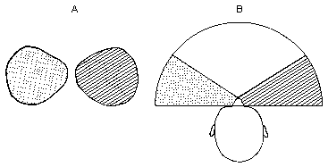
Visual information coming from both eyes is fused in the visual cortex in the back of the brain. The central part of the visual field is seen by both eyes (Fig.22). On both sides of this central, binocular field there are half moon formed parts of visual field that are seen by only one eye.
 |
Fig.22. Visual field. |
We use our peripheral or side vision when moving around. The most central part of the visual field is used in sustained near work, e.g., reading. When the visual field is measured with the clinical instruments these instruments measure what the weakest light is that the eye still can see in different parts of the visual field. A measurement like this gives valuable information on diseases of the visual pathways related to glaucoma or neurologic diseases. It does not give information on how the person sees forms or perceives movement in the different parts of the visual field.
The visual field can change in many ways. Therefore it is often difficult to understand how a visually impaired person sees. If the side parts of the visual field function poorly the person may need to use a white cane in order to move around safely, but he may be able to read without glasses. On the other hand, if the side parts function well and the central field functions poorly, the person may walk like a normally sighted person, but may be able to read only the headings of a newspaper.
Contrast sensitivity
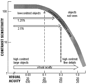
Contrast sensitivity is depicted by a curve (Figure 23A). Under the curve there are the objects that we can see, above and to the right of the slope of the curve is the visual information that we cannot see. Contrast sensitivity can be measured using striped patterns, gratings, or symbols at different contrast levels.
A 
B 
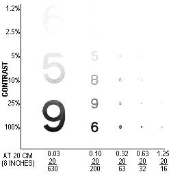
Fig.23. A. Contrast sensitivity curve. B. Visual information at different contrasts in different sizes. Note that large numbers are visible at a fainter contrast than small numbers.
When we measure hearing, an audiogram depicts whichs are the weakest tones at different frequencies that we still can hear. The measurements are made at low, intermediate and high frequencies. When we measure contrast sensitivity we measure what is the faintest grating or symbol still visible when the symbols are large, medium size or small (Fig.23B).
If a visually impaired person has poor contrast sensitivity (s)he cannot see small contrast differences between adjacent surfaces. Everything becomes flat. It is difficult to perceive facial features and expressions. Text in the newspapers seems to have less contrast than before and it is difficult to recognise the edge of the pavement and the stairs.
Contrast sensitivity decreases in several common diseases, diabetes, glaucoma, cataract and diseases of the optic nerve.
Visual adaptation to different luminance levels
A normally sighted person can read by one candle's light and he can read in bright sun light. The difference in the amount of light present in these two situations is million times. The normal person can adapt his/her vision to function at the different luminance levels.
The rod cells of the retina see best in twilight. If they do not function, the person is night blind. Night blindness is the first symptom that develops in many retinal diseases. First the child with a retinal disease starts to see in dim light after an abnormally long waiting. Therefore he will have difficulties in finding his/her clothing in a closet or in a drawer if there is no extra illumination directed into these places. Later (s)he loses night vision completely, even when waiting for a long time (s)he does not start to see in the dark.Changes in visual adaptation time can be easily detected with the CONE Adaptation Test.
Photophobia and delayed adaptation to bright light are often additional symptoms of abnormal visual adaptation. When normally sighted persons enter from a darker room into a bright light, they also see very little for a second, sometimes it even hurts their eyes. They are dazzled. A visually impaired person may be dazzled for a long time. It is possible to decrease the problem by using absorptive glasses and a hat with wide brim or a visor.
Color vision
There are three different types of the retinal cone cells: some cells are most sensitive to red light, other to green light and the third type is most sensitivie to blue light. Also the "normally sighted" individuals may have minor difficulties with color perception. It is often called color blindness but the term is poorly chosen because these persons are not blind, many of them are unaware that they have anything abnormal with their vision. However, if they compare such colors as moss green, snuff brown, dark purple, and dark grey, all these color may look more or less the same. Small deviations from normal affect only some specific working conditions. That is why colour vision is examined at school before students get advice in career planning.
The screening examination uses pseudoisochromatic plates. Most commonly used test is called Ishihara's test. Screening tests are very sensitive and detect even minor deviations from normal color perception. They do not measure the degree of deviation. For the diagnosis of deviant color perception another test is necessary, a quantitative test in form of small caps with color surfaces in all colors of the spectrum. The diagnosis of color deficiency should never be based on a screening test. If a child seems to have any confusion with colours, colour vision should be examined carefully. It can be started with clear basic colours to teach the concepts similar/different in relation to colors, after which quantitative testing is possible.
Young children may train for the quantitative test by playing the Colour Vision Game. Major colour vision deficiencies are revealed already in this game but the diagnose requires proper measurement using pigment tests.
Binocular vision and three dimensional vision
We have two eyes but see only one picture, image. Visual information coming from the two eyes is fused into one image in the visual cortex. Not all normally sighted have binocular vision. They do not use both eyes simultaneously, together. Some persons look alternatingly with their right or left eye. They are usually unaware that they use their eyes separately. It does not disturb them.
Stereovision or three dimensional vision means that we have depth perception in near vision. When we look far away we have another kind of depth perception. We pay attention to the relative size of objects and which object is partially hidden behind another object. The speed of movement with which an object seems to move when we move our head or move around (called parallax) gives us clues on the distance. Therefore persons who do not have stereovision can still assess depth.
Dominant eye
Dominant or leading eye is the eye that we use when we look very carefully at near or at far and can use only one eye. Even when both eyes are used simultaneously one of the eyes is more dominant than the other. We have hand, foot, and eye preference.
Eye motility and its disturbances
Eye movements are usually well controlled. The eyes look at the same object. Eyes turn because of the function of six eye muscles. If one of the eye muscles is paralysed, the eye turns in an abnormal position, the person sees double images (Fig.24).

|
Fig.24. Eye muscles seen from above. The left outer muscle has developed palsy, the left eye turns inward. |
If an eye muscle is not functioning properly the person sees double when trying to look in the direction where the muscle should function. When the eyes are turned in the opposite direction the double image is fused again. The eye with the disturbed motility is covered until the muscle function returns to normal.
Sometimes there is no disturbance in the muscles themselves but the command to turn eyes in a certain direction is not handled normally because of changes in brain function.
Variation in the nature of visual disability
Different visual functions may become impaired independent of each other. Therefore there are many different types of visual impairement and disability. Sometimes a visually impaired person seems to function in a very confusing way. One moment (s)he seems to function like a normally sighted person and in the next moment like a blind person. A visually impaired person seldom pretends to see less than what (s)he actually sees.
One reason for variation in visual behaviour might be changes in illumination. Another may be that (s)he knows the surroundins so there is no difficulty in orientation. Normally sighted move about the same way at home in the dark. They move confidently and securely as long there is nothing unexpected in their way. If somebody leaves an object on the usual path they may trip over it. In the very same way a visually impaired person needs only a few visual cues in a well known place in order to be able to move freely.
If it is difficult to understand how a visually impaired person sees it is quite proper to ask him/her about his/her vision. Most visually impaired people are able to describe the nature of their impairement so well that it is possible to understand their situation better. Some persons say that they have only 10% vision left. Such a number does not describe the degree of visual impairment. The person may be able to move freely relying on his/her vision or may function like a nearly blind. That number (10%) usually means that his/her visual acuity is 20/200 (6/60 or 0.1) and it describes only one of many visual functions.
If the loss of visual functioning is caused by brain damage, the behaviour of the person may look even more perplexing than when the loss is caused by changes in the eyes. In the higher visual functions, perceptual functions, small specific areas of the brain cortex are responsible for specific perceptions. If such an area with specific function is damaged, the corresponding function is either weak or completely lost. Thus an otherwise normally sighted person may not recognize people, not even close relatives. (S)he sees faces but cannot connect the visual information with pictures of faces in his/her memory.
There can be an isolated loss of motion perception, so that the person cannot tell whether a car is moving or not, or in milder cases, may perceive some movement but not how fast the car may be approaching. Colour perception may be disturbed. Recognition of geometric forms may be lost and thus learning letters and numbers may be impossible.
The structure of egocentric space may be lost and thus concepts like 'on the right', 'on the left', 'in the middle', 'next', may be difficult. Also drawing of simple pictures or even copying pictures of angles may be impossible.
It is important that these children/adult persons are not diagnosed as intellectually disabled if they have other functions where they function normally. An uneven profile of functions should always lead to a thorough assessment of all cognitive visual functions and auditory perception. Children with loss of recognition of facial features or facial expressions are sometimes diagnosed as autistic, which is a tragic error and may negatively affect the child's future.