Lateral Geniculate Nucleus
Lateral geniculate nucleus is the large structure of well-organized layers of cells where information from the eyes is relayed to the second neuron of the long retinocalcarine pathway. Visual information undergoes great changes at the level of LGN. There are numerous cell groups in the near-by brain structures (brain stem and thalamus) that affect the function of the LGN neurons. A less known modifier of the inward coming visual information is the information flowing in from the visual cortices to the LGN (top-down).

Figure 5. Simplified picture of the connections that may affect the functions of a LGN cell.
We usually think of the flow of visual information as a flow from the eyes to the brain. At the level of the LGN and in the brain cortex the number of fibres bringing information top-down is much greater than the number of fibres bringing the information toward the cortex. The top-down information affects the in-coming information so that it is reduced and suitable as the information “needed” for the on-going processes in the cortex. Much of the information that comes into the LGN neurons is filtered away. It is estimated that only 20 to 40% of the incoming information is transferred further. If there are losses of cortical functions, then also their effect on the visual information at the level of the LGN is likely to differ from the norm. We have no clinical tests that would characterize possible changes in processing of visual information at the level of the LGN.
From the LGN visual information is transferred further through the optic radiation. This area of brain around the brain ventricles (fluid filled cavities in the middle of the brain) is highly vascular. The vessels may be damaged by circulatory failure during pregnancy or birth or soon after birth. This causes asphyxia/hypoxia, too little oxygen in the surrounding brain structures; haemorrhage in the periventricular structures and even into the ventricles. If the changes are only in the area of the optic radiation, the child may be neurologically normal, yet has losses in a few or several visual functions.
A. |
B. |
|
 |
 |
|
 |
||
C. |
||
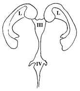
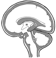
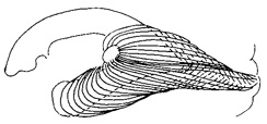
Figure 6. The close anatomic connection between brain ventricles and the optic radiation is seen in these pictures. A. The brain ventricles as seen from the front of the head. B. The fluid filled cavities (marked with dark grey, except the third ventricle and the right lateral ventricle) are both in the middle of the brain and surround it. Lateral ventricles (L) are connected to the fourth ventricle (IV) via the third ventricle (III). In this picture they are seen from the side of the head. C. The nerve fibres of the optic radiation lay next to the wall of the lateral ventricle when they leave the lateral geniculate nucleus.
Visual information flows into the brain through the magnocellular (M) and parvocellular (P) fibres entering different layers in the primary visual cortex. The main components of the information are decoded and encoded in different structures for further processing: 1) colour information in the round structures called “blobs”, 2) contrast edges, line structures in the interblob structures and 3) motion related information in a separate layer. In our clinical examination of vision we usually assess form perception as recognition visual acuity at high contrast only, colour vision more seldom and motion related information not at all. For education and rehabilitation the assessment of quality of vision needs to be much more thorough.
A.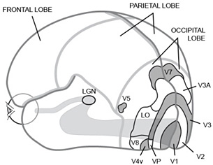
B.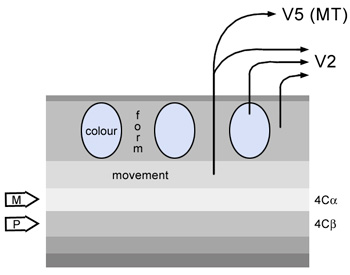
Figure 7. A. Visual pathways from the eye via the LGN to the cortex. B. Structure of the primary visual cortex.
![]()
![]()
![]()
[ Instructions I Paediatric Vision Tests I Vision Tests ]
This document was last modified on