Processing of Visual Information in the Brain
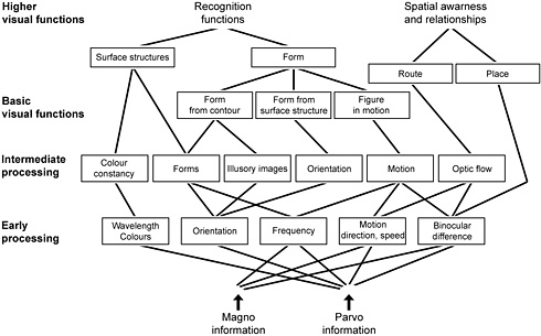
Figure 8. This diagram is a simplified representation of visual pathways in the cortex and covers only visual information that arrives via lateral geniculate nucleus and optic radiation and is processed either in the recognition functions (ventral stream) or in the dorsal stream where awareness of space, orientation in space, eye-hand-coordination are mainly located. Visual information via the tectal pathway is mixed with the more consciously used information from the retinocalcarine pathway in the dorsal stream functions. In a diagram we usually place the different functions in boxes, which makes it easier to see the connections but may give the wrong idea that the functions are in well-defined compartments.
The networks are extremely complex and information flows in both directions, more in the direction top-down, which we often forget. If a child or person has lesions in the higher processes, the top-down information from such an area is less than normal or totally absent, which affects the incoming information. The information that normally is guided to the now affected area is not allowed to enter because “nobody is asking for it”. The child is devoid that information, blind to it. This we often see for example in face blindness, which in young children may look like a total unawareness of faces and pictures of faces but in a few years develops to ability to handle pictures of faces, compare them but still there seems to be no direct connection to memory and thus the child cannot recognise a face as a face of a person although he sees it quite well as a picture.
Visual information goes through such a complex neural network that even the simplest diagrams look difficult to grasp. We do not need to know the details in the physiology of the networks in order to understand unusual behaviours of children with brain damage but it is helpful, if we are aware of the basic facts like 1) there are numerous specialized cortical areas that have specific functions; 2) visual information goes through a decoding and encoding when it arrives into the brain.
The first fact explains how a child/person may have loss of one single visual function, for example does not recognise people by their faces or does not recognise objects by sight but by their haptic form and surface quality (like a blind person). These recognition functions are located in the lower part of the temporal lobe. The loss of function can be also in perception and interpretation of biological movements so that body language cannot be seen or a typical movement pattern of a person cannot be perceived and therefore for example movements in gymnastics must be taught using tactile and kinaesthetic information. In other cases the losses can be vast, for example so that a child is unaware of his body, poorly unaware of the spatial structure of the surrounding space, is without visual memory and visual imagination although other visual functions like visual acuity and visual field are normal. A child may use certain visual information in certain types of functions, for example, in eye-hand coordination the hand functions adequately turning object in correct orientation, yet the child cannot purely visually decide when objects or lines are parallel, in the same direction. The hand receives the information in the parietal lobe and at the same time the purely visual recognition cannot use the information in the temporal lobe.
The second fact explains why a child may have normal form perception in visual acuity tests but cannot perceive fine line structures like threads in materials or draw diagrams on paper with numerous thin lines. This problem can be tested with LEA Grating Acuity test or LEA GRATINGSby asking the child how he sees the lines (which is a difficult task) or asking the child move a finger along the lines with different width (0.5 cpcm to 4 cpcm).
If a child has losses of higher visual functions other than visual acuity, it is common to hear people saying “He sees what he wants to see”. We normally sighted can see what we want to see, can inhibit visual information from entering our attention, stare without using visual information. Brain damage often causes weakness in inhibitory functions. Children cannot prevent surrounding environmental information from entering their consciousness, which makes them hypersensitive to sensory information. At the same time many children have slow shift of attention from one target to the next and may not be able to direct attention toward a part of the visual field, most often to the left half of the visual field. They would really like to see like other children but some of the connections in their brain do not work. Therefore everyone involved in the care and teaching of a child should understand the problems that the child has in using visual information.
![]()
![]()
![]()
[ Instructions I Paediatric Vision Tests I Vision Tests ]
This document was last modified on