Neural Functions in Colour Vision
Perception of colour is based on three different neural functions:- Absorption of light energy in three types of cone cells of the retina;
- Comparison of the absorption rates in these three different cones; and
- Abstraction of colour by cerebral cortex from this comparison.
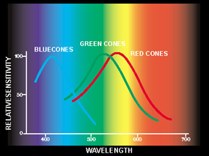
Figure 1 A. Absorption curves of the three cone populations show that each cell absorbs light energy within a wide range of the colour spectrum. Neural impulses coming from all three types of cones selectively activate the cells of the ganglion cell layer from where the impulses are transferred via optic nerves and optic radiations to the primary visual cortex, from where the information moves to colour specific areas for further analysis.
The three types of cone cells are the L-(long wave-length sensitive or "red") cones, the M-(middle wave-length sensitive or "green") cones, and the S-(short wave-length sensitive or "blue") cones. The L- and M-cones constitute the majority of cones, 85 to 90 percent.
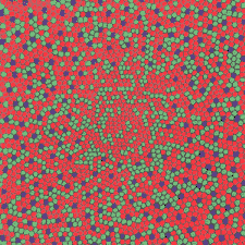
Figure 1 B. The distribution of cone cells in the fovea.
The distribution of cones varies in different locations of the retina: S-cones (“blue cones”) are not present in the very center of the fovea and are concentrated on a circular area, approximately 2° from the center of the fovea.
Differences in cone distribution are probably not important because the integration and comparison of different cone types (i.e., L versus M, and separately, L and M versus S) is probably made on a similar basis in each unit area of colour space. The absence of S-cones in the foveola is probably compensated for within the unit area of colour space that includes the foveola.
Results of colour vision tests vary as a function of the size of the colour stimulus because of the uneven distribution of the cones. In the peripheral retina, we all have "defective" colour perception of small test stimuli. In everyday life, we are not aware of variations in colour perception in the different parts of the visual field, because of the complicated summation functions of the brain.
![]()
![]()
![]()
[ Instructions I Paediatric Vision Tests I Vision Tests ]
This document was last modified on